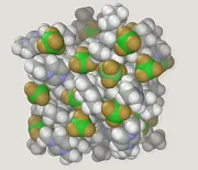
Building the Body Anew: Five Pioneering Biomaterials Transforming Regenerative Medicine
Photo: Distilledwalex (Alabi Olawale), CC BY-SA 4.0, via Wikimedia Commons
The human body is a remarkable self-repairing system — up to a point. Skin heals. Bone knits. Even the liver can regenerate substantial portions of itself under the right conditions. But the body's native repair capacity has hard limits. Severed spinal cord tissue does not reconnect. A failed heart does not grow replacement muscle. Cartilage, once worn away, does not return.
For much of medical history, those limits were simply accepted. Regenerative medicine — the discipline devoted to overcoming them — has existed as a field for several decades, but only in recent years has it acquired the material tools sophisticated enough to match its ambitions. The following five biomaterials represent some of the most consequential advances now moving from scientific discovery toward clinical reality.
1. Decellularized Extracellular Matrix Scaffolds
From cadaver to custom tissue architecture
When a cell is stripped away from its surrounding structural framework, what remains is the extracellular matrix — a three-dimensional scaffold of proteins, glycoproteins, and signaling molecules that once told the cell where it was, what it should do, and how it should grow. Researchers discovered that this matrix, when carefully preserved and decellularized, could serve as a biological blueprint for regeneration.
At the University of Pittsburgh's McGowan Institute for Regenerative Medicine, scientists have used decellularized matrices derived from pig tissue to regenerate muscle in human patients who had lost significant mass to traumatic injury. In published case studies, men who had lost more than 50 percent of a major muscle group regained meaningful functional capacity after implantation — outcomes that would have been inconceivable with conventional surgical repair alone.
The principle works because the preserved matrix retains the precise architectural cues that guide stem cells to differentiate and organize correctly. The body, in effect, reads the scaffold as instructions. Current research is extending this approach to cardiac tissue, tracheal reconstruction, and even partial liver regeneration, with early clinical trials underway at several U.S. academic medical centers.
2. Self-Assembling Peptide Nanofibers
Molecular construction at the nanoscale
Some of the most elegant work in biomaterials science involves molecules that do the engineering themselves. Self-assembling peptide nanofibers are short chains of amino acids designed to spontaneously organize into fibrous networks when introduced into physiological conditions — essentially forming a gel-like scaffold in situ, at the site of injury.
Developed in part through foundational research at MIT by Samuel Stupp and colleagues, these materials have shown particular promise in neural regeneration. When injected into the site of a spinal cord injury in animal models, peptide nanofiber scaffolds created a permissive environment for axonal regrowth — something the body's native inflammatory response actively suppresses. Paralyzed mice regained significant hind-limb motor function in landmark studies published in Science.
Human trials are proceeding cautiously, given the complexity of spinal cord biology, but the underlying principle — using molecular self-assembly to create transient, biodegradable scaffolds that guide tissue repair and then dissolve harmlessly — is being applied to wound healing, bone defect repair, and corneal regeneration with encouraging early results.
3. Bioprinted Hydrogel Constructs
Printing living tissue, one layer at a time
Three-dimensional bioprinting has moved steadily from science fiction toward clinical reality over the past decade. The key enabling material is the bioink: a hydrogel medium — typically derived from alginate, gelatin, or hyaluronic acid — that can carry living cells through a print nozzle without destroying them, then solidify into a defined architecture that supports cell survival and function.
Wake Forest Institute for Regenerative Medicine, led by Anthony Atala, has been among the most prominent American institutions advancing this technology. Researchers there have bioprinted ear cartilage, bone segments, and muscle constructs that, when implanted in animal models, integrated with surrounding tissue and developed functional vascularization. Atala's team has also reported the implantation of bioprinted bladder constructs in human patients — among the earliest examples of a printed organ functioning in a living person.
The current frontier is vascularization at scale: coaxing printed tissue to develop the dense capillary networks required to sustain thicker structures. Several research groups are now incorporating sacrificial channel architectures into their print designs, creating pathways that endothelial cells can colonize to form functional blood vessels.
4. Conductive Polymer Composites for Cardiac Repair
Restoring the heart's electrical conversation
The heart is not merely a pump — it is an electrically coordinated organ whose function depends on the precise propagation of signals across billions of cells. When a myocardial infarction destroys cardiac muscle, the resulting scar tissue is electrically inert, disrupting that coordination and predisposing survivors to arrhythmia and progressive heart failure.
Conductive polymer composites — biomaterials engineered to conduct electrical impulses while remaining mechanically compatible with heart tissue — represent a direct approach to this problem. Materials based on polypyrrole, polyaniline, and graphene-polymer hybrids have been developed that can be fashioned into cardiac patches, applied to infarcted regions, and gradually integrated as the surrounding tissue remodels.
Research teams at institutions including Harvard's Wyss Institute and the University of Washington have demonstrated that conductive patches reduce arrhythmia incidence and preserve ejection fraction in animal models of heart attack. The materials can also be seeded with cardiomyocytes derived from induced pluripotent stem cells, creating patches that are both electrically active and biologically alive. Phase I human safety trials are currently recruiting participants at multiple U.S. sites.
5. Mineral-Organic Composite Bone Substitutes
Engineering bone that thinks it belongs
Bone grafting — transplanting material to fill skeletal defects caused by trauma, tumor resection, or congenital abnormality — has historically relied on autograft tissue harvested from the patient's own body, a procedure that creates a second surgical wound and limited available supply. Synthetic bone substitutes have long existed, but early formulations lacked the biological responsiveness that makes natural bone dynamic and self-renewing.
A newer class of mineral-organic composites addresses this limitation directly. These materials combine hydroxyapatite — the mineral phase of natural bone — with bioactive organic components such as collagen, silk fibroin, or polymer matrices loaded with growth factors. The result is a scaffold that mimics bone's hierarchical structure, degrades at a rate matched to new bone formation, and actively recruits the osteoblasts and osteoclasts responsible for ongoing remodeling.
Researchers at Northwestern University and Columbia University Medical Center have developed variants of these composites that incorporate controlled-release systems for bone morphogenetic proteins, reducing the supraphysiological doses previously required and the associated side effects. Clinical use in craniofacial reconstruction and long-bone defect repair is expanding, with several products having received FDA 510(k) clearance in recent years.
The Road Ahead
Each of these materials represents not a finished product but a point on a trajectory. The shared challenge across all five is translation: moving from promising animal data and early human trials to safe, effective, scalable therapies accessible to patients regardless of geography or economic circumstance. Manufacturing consistency, long-term biocompatibility data, regulatory clarity, and reimbursement structures all remain active areas of work.
What is already clear is that the foundational premise of regenerative medicine — that the body can, with the right materials and signals, rebuild what disease and injury have taken — is no longer aspirational. It is, in growing measure, demonstrable. The science of building the body anew has arrived.